Microscopy
The gold standard in malaria testing is the peripheral smear examination. Thick and thin smears of the blood are both needed for this test. The peripheral blood smear provides comprehensive information on the species, the stages, and the density of parasites with a sensitivity of 5 to 10 parasites/µL of blood for an experienced laboratory professional.
The efficiency of the test depends on the
• quality of the equipment and reagents
• the type and quality of the smear
• skill of the technician
• the parasite density
• and the time spent on reading the smear
The test takes 20 to 60 minutes and is estimated to cost about 12 to 40 US cents per slide in the endemic countries.
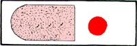
Each slide needs a thick and thin blood sample to examine. Thick smears are therefore used to detect infection, and to estimate parasite concentration.
Alternative Microscopy Methods & RDT
Alternative microscopic methods
These methods have been tried, but inability to easily differentiate the Plasmodium species, requirements of expensive equipment, supplies and special training as well as the high cost limit the use of these methods.
Rapid Diagnostic Tests (RDTs)
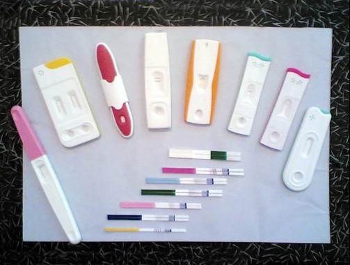
The immunochromatographic tests for the detection of malaria antigens. These tests are widely used for prompt on-site diagnosis in remote endemic areas where reliable microscopy is absent. Aberrant results, whereby negative test results occur at high parasite densities has long been a problem with this form of testing.
Recent studies suggest that false-negative malaria RDT results will rarely occur due to a prozone-like effect in high-density infections, and other causes are more likely. However, RDT line intensity is poorly indicative of parasite density in high-density infections and RDTs should, therefore, not be considered quantitative. Thererfore individuals with suspected severe malaria should rely on peripheral smear testing for accurate parasite counts.